Lower-Leg or Ankle Location
Venous ulcers frequently appear in the lower leg, particularly around the ankle region.

 EN
Phoenix Valley Vein Evaluation for Leg Ulcers
A sore on the lower leg that does not heal normally deserves medical attention. When a wound appears with ankle swelling, skin discoloration, heaviness, itching, hardened skin, or varicose veins, chronic venous disease may be contributing.
✓
Vein consultation at every location
✓
Same-day ultrasound and results when clinically appropriate
✓
Evaluation and vein treatment available across EVC clinics
✓ Vein consultation
✓ Ultrasound when appropriate
Credentials, Professional Affiliations & Training



Understand the Wound
A leg ulcer is an open sore that heals slowly or does not follow the normal healing process. Venous ulcers commonly develop on the lower leg, especially around the ankle, but other ulcer types can look similar and require different care.
1
A persistent wound that does not progress through normal healing deserves medical evaluation.
2
Venous ulcers are often relatively shallow and may have uneven rather than perfectly round borders.
3
Some ulcers release fluid. Changes in drainage amount, color, odor, or consistency can require professional assessment.
4
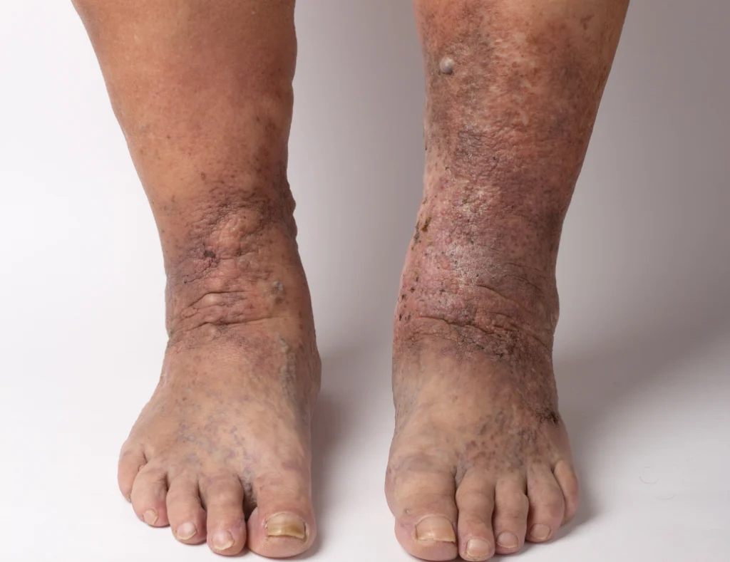
Swelling, itching, discoloration, inflammation, thickening, or hardening may offer clues about the underlying cause.
How Chronic Vein Disease Can Affect the Skin
A venous ulcer can develop when long-term reflux and venous pressure contribute to swelling, inflammation, skin damage, and eventual breakdown. This is a possible progression, not an inevitable outcome.
1
Healthy valves normally help move blood upward through the legs.
2
When valves do not close properly, reflux can allow blood to pool and increase pressure in the lower-leg veins.
3
Long-term venous pressure can contribute to edema, itching, inflammation, and tissue changes.
4
Some people develop reddish-brown pigmentation, stasis dermatitis, or thickened and hardened tissue.
5
With advanced chronic venous disease, weakened skin may eventually open and form a venous ulcer.
Look at the Full Pattern
The wound is only one part of the evaluation. Location, swelling, surrounding skin, visible veins, and symptom history can help determine whether chronic venous disease may be involved.
Venous ulcers frequently appear in the lower leg, particularly around the ankle region.
Chronic venous disease may produce edema around the wound or elsewhere in the lower leg.
Long-term venous pressure may be associated with darker pigmentation in the surrounding tissue.
Venous stasis dermatitis can cause itchy, irritated, flaky, or inflamed skin.
Long-standing inflammation may cause the skin and underlying tissue to become firm or hardened.
Visible veins, aching, heaviness, or a history of venous disease may provide additional clues.
Possible Causes
Not every leg ulcer comes from vein disease. Different wound types can have very different causes, testing needs, and treatment plans.
Faulty vein valves can create sustained venous pressure that contributes to inflammation, skin damage, and venous ulcer formation.
Reduced arterial blood supply can cause wounds that heal poorly and require a different circulation assessment.
Diabetes can impair healing, while reduced sensation may allow pressure or injury to go unnoticed.
Long-term pressure on certain areas can damage skin and underlying tissue, especially when mobility is limited.
Cuts, impacts, surgery, or other injuries may heal poorly when circulation or other medical factors are involved.
Infection, vasculitis, inflammatory disease, and other conditions can also create chronic wounds.
Compare the Clues
Similar-looking wounds can have very different causes. The pattern below helps explain why a clinical and circulation assessment matters before treatment.
Possible Venous Pattern
Often lower leg or ankle, with swelling, brown or reddish skin changes, itching, hardened tissue, or other signs of chronic venous disease.
Arterial Evaluation Needed
Reduced arterial blood supply can create painful, slow-healing wounds, often on the foot, toes, heel, or other distal areas.
Diabetes / Wound Care Evaluation
Neuropathy can reduce sensation and allow pressure injuries to go unnoticed. Diabetes may also slow wound healing.
Know When to Act
A leg ulcer that is not healing, becomes worse, or develops new symptoms should receive medical attention, especially when swelling, skin changes, or visible veins are also present.
Some leg wound symptoms require prompt medical attention rather than waiting for a routine vein appointment.
Book a Vein ConsultationA persistent open wound that is not progressing through normal healing should be evaluated rather than managed indefinitely at home.
Drainage can occur with venous ulcers, but a change in amount, color, odor, or consistency may require professional assessment.
Swelling, brown pigmentation, itching, thickened skin, or visible veins may suggest an underlying venous component.
These changes may indicate infection or worsening inflammation and should receive prompt medical attention.
Fever, chills, worsening redness, increasing pain, or foul-smelling or cloudy drainage are reasons to seek medical care.
Your Vein Evaluation
EVC does not assume that every leg wound is caused by vein disease. The goal is to determine whether the venous circulation may be contributing and whether a separate wound, arterial, diabetic, or infectious evaluation is also needed.
1
Discuss when the wound appeared, how long it has been present, whether it has changed, and any related swelling, drainage, skin changes, or previous wound history.
2
Your provider examines the wound and surrounding skin, along with swelling, visible veins, and other findings that may suggest an underlying venous condition.
3
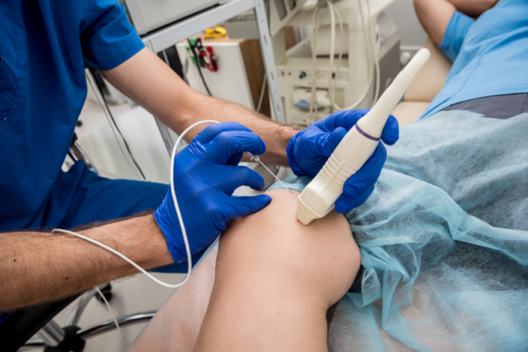
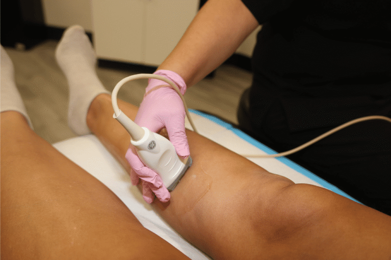
When clinically appropriate, duplex ultrasound assesses blood flow, vein anatomy, valve function, and venous reflux.
4
Your provider explains whether an underlying vein condition may be contributing to the leg ulcer and whether additional wound care or another type of medical evaluation is needed.
Treating the Underlying Vein Condition
Vein treatment and wound treatment are not the same thing. When ultrasound confirms reflux that may be contributing to a venous ulcer, EVC treats the diagnosed vein problem while professional wound care may continue separately.
Catheter-Based
Controlled radiofrequency energy closes selected refluxing superficial veins through a small catheter placed under ultrasound guidance.
✓
Targets selected underlying reflux
✓
Performed in office
✓
Chosen after ultrasound evaluation

Medicated Microfoam
Injectable microfoam closes selected veins affected by venous reflux when the anatomy, symptoms, and treatment plan are appropriate.
✓
Nonthermal treatment option
✓
Useful for selected vein patterns
✓
Chosen after clinical evaluation
Injection Treatment
An injectable sclerosant closes selected abnormal veins and may form part of a broader treatment plan based on the vein pattern identified.
✓
Targets selected smaller veins
✓
May complement a broader plan
✓
Session needs vary by patient
Medical Adhesive
Medical adhesive closes selected diseased superficial veins without thermal energy. Ultrasound findings help determine whether the vein anatomy is appropriate.
✓
Nonthermal closure option
✓
Treats selected refluxing veins
✓
Chosen after ultrasound evaluation
Treating venous reflux can address one contributor to a venous leg ulcer, but ulcer healing also depends on wound care, swelling control, arterial circulation, infection status, and overall health.
The Vein-Treatment Process
Ulcer healing depends on more than a vein procedure. Treating confirmed reflux may address abnormal venous pressure, while the wound itself may still need professional wound care, appropriately selected compression, and follow-up based on the overall medical picture.
01
▤
Confirm the ultrasound findings, affected veins, and why a specific procedure has been selected.
◯
Your care team prepares the leg and explains the procedural steps relevant to the selected treatment.
⌁
Your provider treats the diagnosed vein using the method chosen for your anatomy and venous condition.
02
✓
Vein treatment does not replace professional wound care. Continue the wound, dressing, and compression plan your healthcare team considers appropriate.
↗
Your healthcare team may monitor wound progress, drainage, swelling, pain, skin changes, and other findings over time.
◷
Follow-up helps assess the treated venous disease and whether additional vein care is appropriate.
Understanding Your Benefits
Coverage depends on the underlying vein diagnosis, symptoms, clinical findings, ultrasound results, medical necessity, prior treatment requirements, and the terms of your individual plan. Having an open wound by itself does not automatically determine coverage for a vein procedure.
✓
Elite Vein Clinic works with most major insurance plans
✓
Free insurance benefits verification is available
✓
Our team can explain plan requirements and your estimated financial responsibility
Coverage and patient responsibility vary by plan, diagnosis, treatment, authorization, deductible, coinsurance, and documented medical necessity.
1
Discuss your leg ulcer, related symptoms, insurance information, and possible next steps.
2
Determine whether clinical findings and ultrasound, when appropriate, support a venous diagnosis.
3
Check coverage, documentation, referral, and authorization requirements for the diagnosed vein condition.
4
Understand known plan requirements and estimated financial responsibility before treatment.
Why Choose Elite Vein Clinic?
A wound can have several possible causes. EVC evaluates whether the venous circulation may be contributing.
When the pattern suggests a vein problem, on-site ultrasound can identify reflux and other venous abnormalities.
EVC offers RFA, Varithena, compounded sclerotherapy, and VenaSeal for selected venous conditions.
A vein procedure is recommended only when the evaluation identifies an appropriate venous problem.

Your Vein Care Team
Learn about the physicians who assess venous disease and guide personalized treatment across Elite Vein Clinic locations.


Lead Practitioner
Board-Certified Emergency Medicine
Ultrasound-Guided Procedures

Vein Specialist
Dr. Olaide Abiodun is a physician specializing in phlebology and the diagnosis and treatment of venous disease. She cares for patients with varicose veins, chronic venous insufficiency, leg swelling, heaviness, aching, and other symptoms related to poor venous circulation.
She is board-certified in Internal Medicine and brings more than a decade of clinical experience treating complex medical conditions across hospitals in Ohio and Arizona. Her broad medical background provides a strong foundation for evaluating patients with underlying health conditions that may contribute to or complicate chronic vein disease.
At Elite Vein Clinic, Dr. Abiodun focuses on personalized, minimally invasive vein treatments designed to address the underlying source of venous symptoms while helping patients improve comfort, mobility, and overall quality of life.
Known for her compassionate approach, Dr. Abiodun combines advanced phlebology care with individualized treatment planning and patient education to help each patient feel informed and confident throughout their treatment.
Book Consultation
Lead Practitioner
Dr. Jaime French is a physician specializing in phlebology and the diagnosis and treatment of venous disease. She provides care for patients with varicose veins, chronic venous insufficiency, leg swelling, heaviness, aching, and other symptoms related to poor venous circulation.
Dr. French is board-certified in Emergency Medicine and earned her medical degree from Midwestern University’s Arizona College of Osteopathic Medicine before completing her residency at the University of Arizona in Tucson. She later practiced at HonorHealth Emergency Departments throughout the Valley, where she developed extensive experience with ultrasound-guided procedures, vascular evaluation, and complex medical conditions.
At Elite Vein Clinic, Dr. French focuses on minimally invasive, image-guided vein treatments designed to address the underlying cause of venous disease while allowing patients to return to normal activity with minimal downtime.
Known for her thoughtful and approachable style, Dr. French combines advanced phlebology care with individualized treatment planning and patient education to help improve leg comfort, mobility, appearance, and overall quality of life.
Book ConsultationPatient Experiences
Read experiences from patients who visited Elite Vein Clinic for evaluation and vein care.


Common Questions
Clear answers can help you understand ulcer types, the role of venous disease, and when vein evaluation or other medical care may be appropriate.
What is a leg ulcer?
A leg ulcer is an open sore that does not heal normally. Leg ulcers can have venous, arterial, diabetic, pressure-related, traumatic, inflammatory, infectious, and other causes.
What is a venous leg ulcer?
A venous leg ulcer is an open wound associated with chronic venous disease and persistently elevated pressure in the leg veins. It commonly develops on the lower leg or around the ankle.
What are the early signs of a venous leg ulcer?
Some people develop swelling, heaviness, itching, reddish-brown discoloration, stasis dermatitis, or hardened skin before ulceration. These changes do not guarantee that an ulcer will develop.
What does a venous ulcer look like?
Venous ulcers are often shallow wounds with irregular borders. Surrounding skin may be swollen, discolored, irritated, shiny, tight, or hardened. Appearance alone cannot confirm the diagnosis.
Where do venous leg ulcers usually develop?
They commonly occur in the lower leg, particularly around the ankle region.
Are all leg ulcers caused by poor veins?
No. Arterial disease, diabetes, neuropathy, pressure, injury, infection, inflammatory conditions, and other problems can also cause chronic wounds.
What is the difference between a venous and arterial ulcer?
Venous ulcers are associated with abnormal venous pressure, while arterial ulcers occur when insufficient arterial blood reaches tissue. Because their causes differ, their evaluation and treatment differ as well.
Can diabetes cause leg ulcers?
Yes. Diabetes can impair wound healing and may cause neuropathy, making injuries harder to notice. Diabetes and arterial circulation problems may also occur together.
Does a leg ulcer need a vein ultrasound?
Not every ulcer does. When the wound pattern and accompanying symptoms suggest venous disease, duplex ultrasound can help assess venous blood flow, anatomy, and valve function.
Do I need an arterial circulation test too?
That depends on the clinical situation. Arterial circulation often needs consideration before compression therapy for a suspected venous ulcer, especially when arterial disease is possible.
Should I use compression stockings if I have a leg ulcer?
Do not assume compression is appropriate simply from the appearance of a wound. Compression can be important in appropriately diagnosed venous ulcer care, but circulation and the overall condition should first be assessed by a qualified healthcare professional.
Can vein treatment help a venous ulcer?
When venous reflux is contributing, treatment of appropriate refluxing veins may form part of the overall management plan. The wound itself may still require professional wound care and appropriately selected compression.
Will treating my veins make the ulcer heal immediately?
No result should be guaranteed. Healing depends on factors including wound severity, venous pressure, arterial circulation, infection, swelling, diabetes or other medical conditions, and appropriate wound management.
What signs may mean a leg ulcer is infected?
Increasing pain, warmth, redness or swelling, cloudy or foul-smelling drainage, fever, chills, or worsening symptoms can indicate infection and require medical attention.
Which vein treatments does Elite Vein Clinic offer?
EVC offers radiofrequency ablation, Varithena, compounded sclerotherapy, and VenaSeal for selected venous conditions. The appropriate option depends on the diagnosis and ultrasound findings.
English English  Spanish


